
As researchers, most of us currently perform Western Blots and PCR, but working in the field of epigenetics, we can use other techniques.
Immunofluorescence is an immunohistochemistry technique that uses fluorophores to visualize cellular antigens, such as proteins.
This technique can be utilized to visualize the localization of various cellular components within cells and within cell compartments.
In epigenetics, it’s largely used to observe the distribution of different histone modifications or histone variants and so to have an idea of the nuclear organization and chromatin landscape.
For instance, this allows us to see heterochromatic and euchromatic regions within the nucleus and how their distribution changes in different conditions.
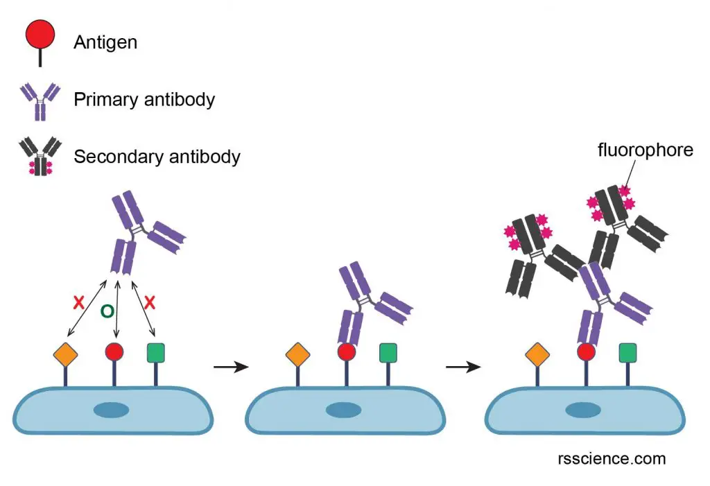
The key steps of the protocol are:
-
- Fixation with formaldehyde, to prevent the degradation of your sample
- Incubation with a primary antibody that specifically recognizes the protein of interest or the PTM of interest
- Incubation with a secondary antibody coupled to a fluorophore and, if your protein interacts with DNA don’t forget to stain your sample with DAPI!
- Observation with a fluorescence microscope or a confocal microscope
Text by Stefania Paltrinieri, PhD Student EpiSeedLink Marie Skłodowska-Curie Actions
Frontal image by Nicola Ferrari